 ShareCompartir

Monthy Case Studies - 2001
Case #63 - July, 2001
A family from Afghanistan immigrated to the United States to live in Missouri. Stool specimens were collected from all family members and submitted to the Missouri State Health Laboratory for an O & P (ova and parasites) examination. The specimens were divided into vials containing 10% formalin and PVA. Smears were made from the PVA preserved specimens, stained with trichrome stain, and examined. The 10% formalin-preserved specimens were concentrated using the FEA (formalin-ethyl acetate) concentration method. Wet mounts of the sediment were examined. Some of the organisms identified in the stool specimens from family members included Giardia duodenalis cysts, Endolimax nana cysts and trophozoites, Hymenolepis nana eggs, and Entamoeba hartmanni cysts and trophozoites. A worm-like object was discovered in the stool concentrate from a 5-year-old family member. Figures A, B, and C are detailed images of the object. What is your diagnosis? Based on what criteria?
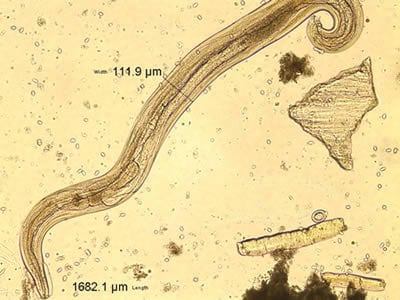
Figure A
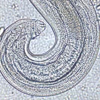
Figure B
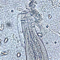
Figure C
Acknowledgement: The images and clinical history for this case were kindly provided by the Missouri State Health Laboratory.
Answer to Case #63
The object shown in the images was a male pinworm (Enterobius vermicularis). Diagnostic features observed were:
- the presence of cephalic alae at the anterior end.
- the presence of a single spicule, indicating that this is a male worm (Ascaris lumbricoides and most other nematodes have two spicules).
- a size range from two to five millimeters in length by 0.1 to 0.2 mm wide. The worm may have shrunk from being preserved in 10% formalin or may not be fully mature.
More on: Enterobiasis (Pinworm Infection)
Images presented in the monthly case studies are from specimens submitted for diagnosis or archiving. On rare occasions, clinical histories given may be partly fictitious.
