 ShareCompartir

Monthy Case Studies - 2001
Case #58 - April, 2001
Laboratorians in the Division of Parasitic Diseases at CDC examined a stool specimen from an HIV-positive patient. Examination of the stool specimen included a formalin-ethyl acetate (FEA) concentration and smears stained with trichrome. Figures A, B, and C show cysts and Figures D, E, and F show trophozoites that were found on the trichrome stained smear. What is your diagnosis? Based on what criteria?
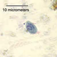
Figure A
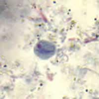
Figure B
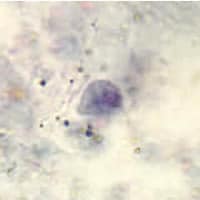
Figure C
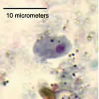
Figure D
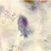
Figure E
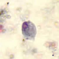
Figure F
Answer to Case #58
The organism shown was Retortamonas intestinalis, a flagellate that has been found in the intestinal tract of humans. Diagnostic features observed were:
- ovoid or pyriform cysts (Figures A-D) ranging from five to seven micrometers in diameter. Cysts of Chilomastix mesnili have a similar morphology, but are slightly larger (seven to ten micrometers).
- uninucleate cysts with a compact central karyosome and varying amounts of peripheral chromatin.
- presence of fibrils (associated with the cytostome) within some of the cysts.
- ovoid or pyriform trophozoites (Figures E and F) within the normal size range for R. intestinalis (5-10 micrometers long). Trophozoites of C. mesnili are normally 10 to 15 micrometers in length.
- the presence of a cytostome at the anterior end that can extend one half the length of the organism.
More on: Retortamonas intestinalis
Images presented in the monthly case studies are from specimens submitted for diagnosis or archiving. On rare occasions, clinical histories given may be partly fictitious.
