 ShareCompartir

Monthy Case Studies - 2000
Case #50 - December, 2000
A 45-year-old woman immigrated to Oregon from Russia four years ago. She was seen in a health care facility with complaints of abdominal discomfort and intermittent diarrhea. A stool specimen was obtained and processed for an O & P (ova and parasites) examination. The objects seen in Figures A through D were seen in her specimen. The objects measured 71 to 85 micrometers in length. What is your diagnosis? Based on what criteria?
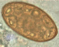
Figure A
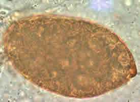
Figure B
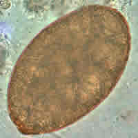
Figure C
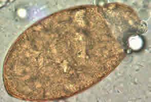
Figure D
Answer to Case #50
This was a case of nanophyetiasis caused by Nanophyetus sp. It was unclear if the infection was acquired in Oregon (in which case it would probably be N. salmincola) or in Russia (in which case it could be N. salmincola or N. schikhobalowi). Diagnostic features observed were:
- operculated eggs that were light brown in color and broadly ovoid. The eggs had a bluntly pointed, thickened, and darkened area of the shell at the abopercular end.
- eggs within the size range for Nanophyetus spp. (64 to 97 micrometers in length by 34 to 55 micrometers in width).
When passed in the feces, Nanophyetus eggs are usually not embryonated. However, if the specimen is not preserved properly, some development can occur, as shown in this case. Distinguishing Nanophyetus eggs from those of Diphyllobothrium latum can be a challenge since they are morphologically similar. Diphyllobothrium latum eggs are a little smaller, ranging in size from 58 to 75 micrometers in length by 40 to 50 micrometers in width. Also, D. latum eggs appear to be broader in relative width than those of Nanophyetus and usually, though not always, have a small knob at the abopercular end. The eggs shown in this case were somewhat larger and narrower than D. latum eggs and they did not possess a knob opposite the operculum. The thickening of the abopercular end of the eggs shown in Figures B and C could be mistaken for a knob you might see in D. latum; however, the thickening is too broad to be called a knob.
Images presented in the monthly case studies are from specimens submitted for diagnosis or archiving. On rare occasions, clinical histories given may be partly fictitious.
