 ShareCompartir

Monthy Case Studies - 2000
Case #48 - November, 2000
A 38-year-old HIV-positive man was seen at a local AIDS clinic for chronic cough and hemoptysis (blood in sputum). A bronchoalveolar lavage (BAL) was performed and an object believed to be nematode larva was observed in one of the stained smears of the BAL. A single object was seen and is shown below at 100×, 400×, and 1000× magnification. The object measured approximately 440 micrometers in length by 30 micrometers in maximum diameter. What is your diagnosis? Based on what criteria?
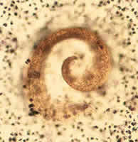
Figure A
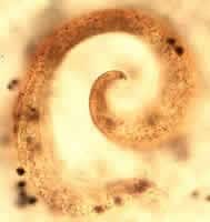
Figure B
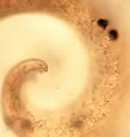
Figure C
Answer to Case #48
This was an artifact that resembled a nematode larva. The artifact was initially considered to be a first-stage Strongyloides stercoralis larva. Several morphologic features indicated this was not a Strongyloides larva. The size and general shape of the structure was not consistent with a parasitic nematode larva. First-stage (rhabditiform) Strongyloides larvae measure 200 to 400 micrometers in length by 15 to 20 micrometers in diameter, while third-stage (infective, filariform) larvae measure 500 to 600 micrometers in length by 15 to 16 micrometers in diameter. This object was slightly longer than the maximum for first-stage larvae and shorter than third-stage larvae. Additionally, the diameter was considerably larger than Strongyloides larvae of either stage. First-stage larvae of Strongyloides have a long, tapered tail, whereas this object had a very short blunt end by comparison. Although some very small, fine particulate matter is visible in the object, it does not resemble typical nuclei seen in stained nematode larvae, and the object appears to be homogenous in appearance and lacks any internal structure.
Several images of Strongyloides larvae are shown below for comparison to the artifact. Figure D below is from a wet mount of stool with a first-stage larva showing the general larval features (captured using DIC); Figure E below shows a stained filariform larva found in a sputum sample; and Figure F below illustrates a rhabditiform (first-stage) larva in a trichrome stained stool smear. Although few internal features are evident in these stained larvae, the general shape of the larvae is evident, and clearly distinct from the artifact presented in this case. One must remember that in disseminated Strongyloides cases, in addition to both first- and third-stage larvae, adult female worms, or even eggs, might also be found.
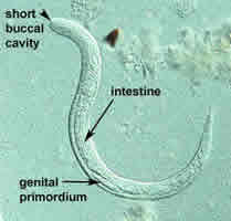
Figure D
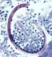
Figure E
Figure F
More on: Artifacts
Images presented in the monthly case studies are from specimens submitted for diagnosis or archiving. On rare occasions, clinical histories given may be partly fictitious.
