 ShareCompartir

Monthy Case Studies - 2000
Case #40 - July, 2000
A 5-year-old boy from Bolivia was seen by a pediatrician at a children's health community clinic in Miami, Florida as part of a preadoption physical examination. No medical history was available at the time of the visit. The doctor ordered stool and blood exams to be performed. No parasites were reported from blood films. Figure A (200×) shows what was seen on a wet mount made from the sediment of an FEA concentration procedure. What is your diagnosis? Based on what criteria?
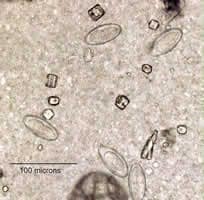
Figure A
Answer to Case #40
This was a case of enterobiasis, caused by Enterobius vermicularis (commonly known as pinworm infection). The objects in the Figure A are eggs of Enterobius vermicularis. Diagnostic features observed were:
- the size of the objects, which was consistent with pinworm eggs (50 to 60 micrometers in length by 20 to 30 micrometers wide).
- partially-embryonated eggs with a shell was thick and colorless, and slightly flattened on one side.
Pinworm eggs are not usually seen in routine stool exams and, if detected in such an exam, often do not appear embryonated.
The diagnosis was compounded by several factors. First, as noted in the case description, this was formalin-fixed stool. Typically pinworm eggs are observed in an unfixed state. Fixation tends to cause cytoplasmic contraction, and thus, there is some distortion of the internal anatomy of the eggs. The fact that all the eggs measure at or near the maximum stated size for pinworm eggs also raised questions about the identification. The fact that the eggs were lying in various orientations and did not clearly show a flattened side also posed a challenge. This case highlights the fact that when a common parasite is seen in an unusual location, it can lead to confusion. We have included additional images of two eggs (500× oil) below that more clearly illustrate the typical features of unembryonated pinworm eggs. These eggs measured 60 micrometers in length.
The most reliable method for diagnosing pinworm infection is the use of pinworm paddles—small, disposable spatulas with adhesive material on one side. The sticky side is applied to the perianal region of the patient early in the morning before bathing or defecating. The spatula is examined, adhesive side up, microscopically for eggs. Pinworm eggs detected using this method are generally embryonated.
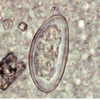
Figure B
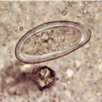
Figure C
More on: Enterobiasis (Pinworm Infection)
Images presented in the monthly case studies are from specimens submitted for diagnosis or archiving. On rare occasions, clinical histories given may be partly fictitious.
