 ShareCompartir

Monthy Case Studies - 2000
Case #30 - February, 2000
Two sisters in their early twenties from Dallas, Texas, went on a ten-day ecotour in Costa Rica. They camped in a tent during most of the trip. The sisters complained of being bitten by various insects during the entire trip. Two months after returning from the excursion, both women noted several small, ulcerated sores on their wrists and necks. They went to a local dermatologist who examined their sores. The physician made touch-prep impression smears from sores of each patient's wrist by pressing a 1" × 5" glass slide against the sores. The images below show impressions from each woman (Figures A and B, Giemsa stain). The physician also took a few small biopsies from the base of the sores on each woman's wrists and neck. Some of the tissue was sent to the pathology lab where it was sectioned and stained with H & E (hematoxylin and eosin), see Figures C and D. The remaining specimens were individually placed in sterile saline and then refrigerated at 4°C. What is your diagnosis? Based on what criteria?
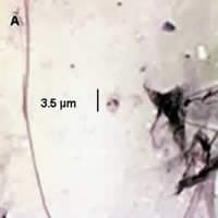
Figure A
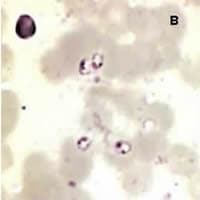
Figure B
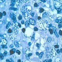
Figure C
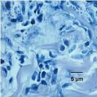
Figure D
Answer to Case #30
This was a case of leishmaniasis caused by Leishmania sp. Morphologically, the amastigotes of the different species of Leishmania are indistinguishable from one another. Leishmania is an intracellular parasitic organism of macrophages. However, they may be observed extracellularly on various slide preparations. The characteristic size (two to four micrometers) and shape (round to oval), as well as internal structures (nucleus and rod-shaped kinetoplast) of Leishmania are used to differentiate it from similar appearing organisms such as Histoplasma sp. The images below show structures consistent with Leishmania sp. (Figures C and D; 1000×, H & E stain). In the images from the tissue sections (Figure C), amastigotes are visible in the clear spaces (red arrows) as well as in the tissue (yellow arrow, Figure D).
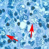
Figure C
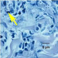
Figure D
More on: Leishmaniasis
Images presented in the monthly case studies are from specimens submitted for diagnosis or archiving. On rare occasions, clinical histories given may be partly fictitious.
