 ShareCompartir

Monthy Case Studies - 1999
Case #22 - October, 1999
A 19-year-old woman, who had lived most of her life in southeastern Alaska, spent a few days on a farm in Washington state during the early summer of 1998. The farm had horses, goats, and dogs. She developed right upper quadrant pain and vomiting on July 19, 1999. Cysts were surgically removed on August 2, 1999 from her liver. Examination of fluid in the cysts revealed the objects shown in the images below (200×). What is your diagnosis? Based on what criteria?
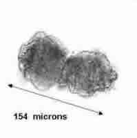
Figure A
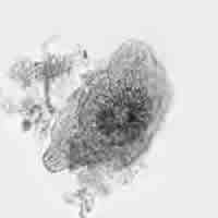
Figure B
Answer to Case #22
This was a tapeworm infection caused by Echinococcus granulosus. This case represents one of the more serious infections caused by larval tapeworms in humans. The diagnostic feature seen in the images were the rostellar hooklets. Figure B is a view of a "squashed" organism as you look down at the rostellum. We did not state that the cysts removed were alveolar cysts. If we had, that would indicate an infection with E. multilocularis.
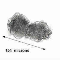
Figure A
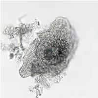
Figure B
More on: Echinococcosis
Images presented in the monthly case studies are from specimens submitted for diagnosis or archiving. On rare occasions, clinical histories given may be partly fictitious.
