 ShareCompartir

Monthy Case Studies - 1999
Case #18 - August, 1999
An HIV-positive man was seen at a local hospital for severe diarrhea. His formalin preserved fecal specimen was stained and the objects shown in Figure A were see at 1000× under oil. What is your diagnosis? Based on what criteria?
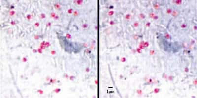
Figure A
Answer to Case #18
This was a case of microsporidiosis. Diagnostic features observed included:
- the size (approximately one micrometer).
- characteristic morphology (round to slightly oval with some spores exhibiting an equatorial band or belt-like stripe). The stain used on the smear for the images was Weber's Chromotrope 2R stain (Weber R, Bryan RT, Owen RL, Wilcox CM, Gorelkin L, Visvesvara GS. Improved light-microscopical detection of microsporidia spores in stool and duodenal aspirates. The Enteric Opportunistic Infections Working Group. N Eng J Med 1992;326:161-166.).
There are two microsporidian species related to intestinal infections in HIV-positive individuals. Enterocytozoon bieneusi is the most prevalent. The other species is Encephalitozoon intestinalis and is usually a little more elongated than E. bieneusi as well as larger (E. bieneusi being an average of one micrometer and E. intestinalis being 1.5 to 2 micrometers). One might say this is possibly an infection with E. bieneusi based on the two criteria above. However, it should only be reported as microsporidia. Species identification of microsporidia can be achieved through electron microscopy, or through immunologic or molecular methods. In this particular case, both immunofluorescence (IF) microscopy and PCR were performed. The IF procedure used detects E. intestinalis (Moura H, Sodre FC, Bornay-Llinares FJ, Leitch GJ, Navin T, Wahlquist S, et al. Detection by an Immunofluorescence Test of Encephalitozoon intestinalis Spores in Routinely Formalin-Fixed Stool Samples Stored at Room Temperature. J Clin Microbiol 1999;37:2317-22.), and in this case the results were negative. The PCR was performed using diagnostic primers as described in the literature (da Silva AJ, Schwartz DA, Visvesvara GS, de Moura H, Slemenda SB, Pieniazek NJ. Sensitive PCR diagnosis of Infections by Enterocytozoon bieneusi (microsporidia) using primers based on the region coding for small-subunit rRNA. J Clin Microbiol 1996;34:986-987.), and in this case E. bieneusi was detected. Thus, the species identification in this infection is Enterocytozoon bieneusi.
More on: Microsporidiosis
Images presented in the monthly case studies are from specimens submitted for diagnosis or archiving. On rare occasions, clinical histories given may be partly fictitious.
