 ShareCompartir

Monthy Case Studies - 1999
Case #16 - July, 1999
A 76-year-old man was admitted to a hospital in Georgia with diabetes, chronic diarrhea, and weight loss. The pathology department sent tissue sections from gastrointestinal biopsies to CDC for diagnostic confirmation. The diagnosis was chronic duodenitis and cross-section of adult or larval round worm. The images are from biopsies of the distal duodenum, focusing on the cross-section of the suspected worm. Figure A was taken at a magnification of 50× (scale bar = 200 micrometers) and Figure B is a close-up, taken at 200× (scale bar = 50 micrometers). What is your diagnosis? Based on what criteria?
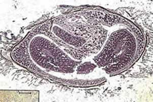
Figure A
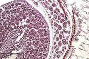
Figure B
Answer to Case #16
The object shown in this case was an artifact (plant material). The submitting laboratory originally suspected that the object might be a cross-sectioned Strongyloides. The object measured about 2.4 by 1.3 mm in diameter, which is too large for Strongyloides. An adult female measures no more than 30 to 40 micrometers (0.03 to 0.04 mm) in diameter. Of the common human nematodes, only Ascaris, which can be up to several millimeters in diameter, are the size of this object. Also, there was no typical body wall composed of a cuticle with underlying muscle cells; no internal tubes (a digestive and one or more reproductive tubes); and no well defined body cavity, as would be typical of nematodes. Figure C below shows a cross section of a nonhuman roundworm trapped in muscle tissue; long axis of section is 950 micrometers. The object also had many individual compartments which is very typical of plant matter. Figure B clearly showed the compartmentalization. The material in the compartments did not bear any resemblance to the reproductive structures seen in nematodes, such as eggs or larvae. The heavy, cellular composition of the outer wall was not typical of a nematode cuticle, but of plant cells.
This object can also be distinguished from trematodes and tapeworms. Many of the same features that distinguish this object from nematodes also distinguish it from flatworms, including the heavy cellular outer coating, the absence of muscle fibers, and the absence of a parenchymous matrix filling the body cavity. The absence of reproductive and digestive tubes, suckers, and other morphologic features consistent with parasites further help identify this as an artifact.
The object is not parasitic in origin, but represents a plant seed or burr of some type. Objects very similar to this have been seen in tissue sections of appendix, throat, and lungs. Whether they are common objects in food, or items that are accidentally ingested is not known. What is clear, is that they can, on occasion, become embedded in various host tissues and cause confusion because they superficially resemble parasites.

Figure A
More on: Strongyloidiasis
Images presented in the monthly case studies are from specimens submitted for diagnosis or archiving. On rare occasions, clinical histories given may be partly fictitious.
