 ShareCompartir

Monthy Case Studies - 1999
Case #9 - April, 1999
A 36-year-old woman with a history of travel to Puerto Rico submitted a routine stool specimen. The images below were captured during wet mount examination of the concentrate from a stool specimen. The images are not on the same scale (see below). What is your diagnosis? Based on what criteria?
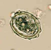
Figure A: 60 µm by 49 µm
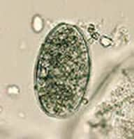
Figure B: 71 µm by 44 µm
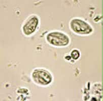
Figure C: 12 µm by 14 µm
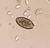
Figure D: 55 µm by 27 µm
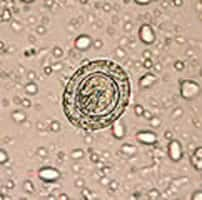
Figure E: 50 µm by 44 µm
Acknowledgement: The images were kindly shared by Dr. Ray Kaplan, Atlanta.
Answer to Case #9
This case demonstrated a mixed infection of ascariasis, caused by Ascaris lumbricoides, giardiasis, caused by Giardia duodenalis, trichuriasis, caused by Trichuris trichiura, and hymenolepiasis, caused by Hymenolepis nana. Diagnostic features included:
- Ascaris lumbricoides eggs (Figures A and B). The egg in Figure A was partially decorticated, with only a few mammillations observable on the outer layer of the shell. It was fertilized, as evidenced by its thick shell, and its structured inner contents are separated from the shell by a space. The egg in Figure B was not fertilized. Compared to the fertilized egg in Figure A, it was more elongated, its shell was thinner, and its inner contents were not structured, but appear globular, and filled the shell, leaving no space. The imperfection on the upper right corner of the shell was somewhat misleading and suggested an operculum; however, careful examination showed that the shell was intact and that the outer covering was all that was missing.
- Giardia duodenalis cysts (Figure C), recognizable by their size, shape, and appearance. Since this wet mount preparation was not stained with iodine, the typical internal structures (axoneme, fibrils, nuclei) were not perfectly defined, but they were partially recognizable. You probably noticed the cysts in the background of the other lower magnification images—particularly in Figure D; even though they were out of focus, you may have guessed that they were Giardia cysts.
- an egg of Trichuris trichiura (Figure D) showing the typical barrel shape, polar prominences or plugs, and a thick shell.
- an egg of the dwarf tapeworm, Hymenolepis nana (Figure E). Visible diagnostic features included: presence of hooklets (three of the six hooklets of the oncosphere can be seen), and size (50 micrometers by 44 micrometers, within the range for H. nana, which is 40 to 60 micrometers by 30 to 50 micrometers). The size distinguishes it from the larger H. diminuta (rat tapeworm) (70 to 86 micrometers by 60 to 80 micrometers). Another diagnostic feature, not observable in Figure E, would be the presence polar filaments between the oncosphere and the shell in H. nana. Such polar filaments are absent in H. diminuta. The image below (Figure F) shows these filaments (at the upper left and lower right corners) in an H. nana egg.
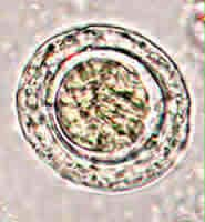
Figure F
More on: Giardiasis
More on: Ascariasis
More on: Trichuriasis
More on: Hymenolepiasis
Images presented in the monthly case studies are from specimens submitted for diagnosis or archiving. On rare occasions, clinical histories given may be partly fictitious.
