
Loiasis
[Loa loa]
Causal Agent
Loa loa, a filarid nematode commonly referred to as the African eye worm.
Life Cycle
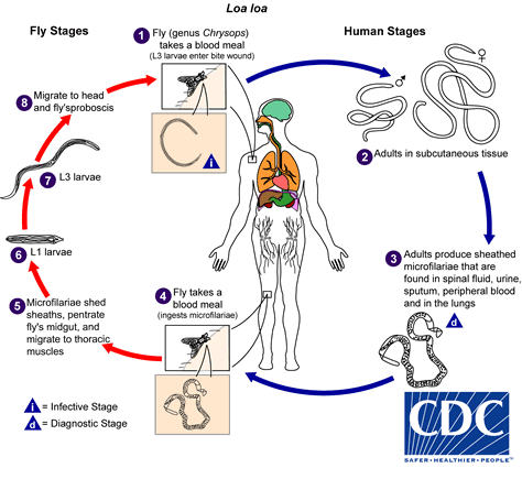
The vector for Loa loa filariasis are flies from two species of the genus Chrysops, C. silacea and C. dimidiata. During a blood meal, an infected fly (genus Chrysops, day-biting flies) introduces third-stage filarial larvae onto the skin of the human host, where they penetrate into the bite wound  . The larvae develop into adults that commonly reside in subcutaneous tissue
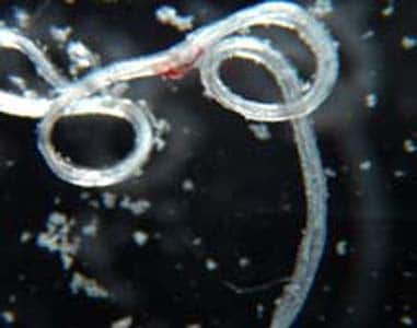
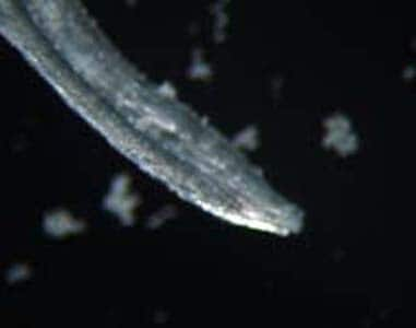
  . The female worms measure 40 to 70 mm in length and 0.5 mm in diameter, while the males measure 30 to 34 mm in length and 0.35 to 0.43 mm in diameter. Adults produce microfilariae measuring 250 to 300 µm by 6 to 8 μm, which are sheathed and have diurnal periodicity. Microfilariae have been recovered from spinal fluids, urine, and sputum. During the day they are found in peripheral blood, but during the noncirculation phase, they are found in the lungs
  . The fly ingests microfilariae during a blood meal
  . After ingestion, the microfilariae lose their sheaths and migrate from the fly’s midgut through the hemocoel to the thoracic muscles of the arthropod
  . There the microfilariae develop into first-stage larvae
  and subsequently into third-stage infective larvae
  . The third-stage infective larvae migrate to the fly’s proboscis
  and can infect another human when the fly takes a blood meal .
Geographic Distribution
Loa loa is found in Africa.
Clinical Presentation
Loiasis is often asymptomatic. Episodic angioedema (Calabar swellings) and subconjunctival migration of an adult worm can occur.
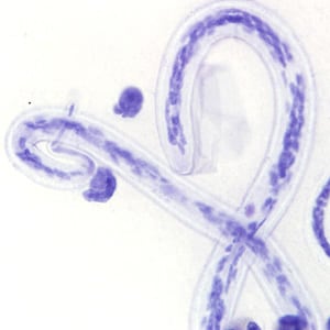
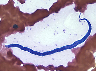
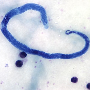
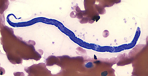
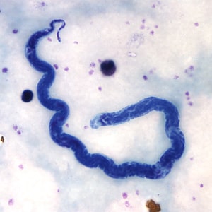
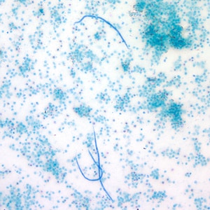
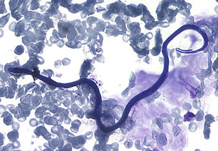
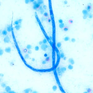
Microfilariae of Loa loa.
Adults of L. loa.
Diagnostic Findings
Loa loa is usually diagnosed by the finding of microfilaria in peripheral blood smears or adults in the subconjunctiva. The blood films may be thick or thin and stained with Giemsa or hematoxylin-and-eosin. For increased sensitivity, concentration techniques can be used. These include centrifugation of the blood sample lysed in 2% formalin (Knott’s technique), or filtration through a Nucleopore® membrane. Microfilariae of L. loa exhibit diurnal periodicity and a diagnosis is best made from blood collected during the mid-day (10 AM-2 PM). The presence of Calabar swellings can aid in the diagnosis.
Treatment Information
Treatment information for loiasis can be found at: https://www.cdc.gov/parasites/loiasis/health_professionals/index.html
DPDx is an educational resource designed for health professionals and laboratory scientists. For an overview including prevention, control, and treatment visit www.cdc.gov/parasites/.